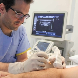
Autologous Blood Injection (ABI) harnesses the healing properties of blood in order to reliably treat pain arising from tendons, ligaments and muscles.
With prolonged exercise and increasing age, pain arising from tendons is becoming more prevalent, particularly in active ‘baby boomers’.
Prior to any proposed ABI, an accurate diagnosis must be made, which usually requires a test such as an MRI or ultrasound to confirm that the specific tendon, ligament or muscle is the source of the patient’s pain. Once referred for an ABI, the radiologist at Melbourne Radiology Clinic will review the patient and discuss the ABI procedure and rehabilitation.
Treatment of Tendinosis
ABI is used for the treatment of tendinosis, also known as tendinopathy, which is the medical term for the more commonly used term 'tendinitis'
With increasing severity of tendinosis, partial thickness tears may form, which if left untreated can result in a full thickness tendon tear. The tendinosis-tear process is simply an increasing spectrum of injury to the tendon. Any tendon can be treated with this procedure and though not used routinely, the procedure may also be used in muscle and ligament tears (‘strains and sprains’).
Ultrasound Guided
Procedure.
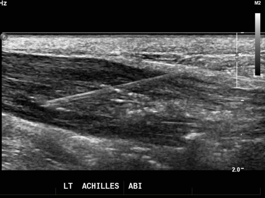
The procedure of ABI involves withdrawing whole blood from the patient, usually taken from the patient’s elbow or forearm, and then injecting it into the area of maximal abnormality using ultrasound.
Ultrasound is used to ensure that the blood is delivered precisely and safely to the area concerned. Platelets, small cells found in blood which are involved in clotting, contain ‘alpha granules’ which release substances such as platelet derived growth factor (PDGF) into the tendon and commence a cascade of natural healing.
Though dependent on the severity of the underlying tendon disease and the duration of symptoms, approximately 80% of patients will obtain complete or significant relief of their symptoms.
Patellar tendinosis.
Inflammation of the patellar tendon generally occurs as a result of repetitive trauma, eg jumping sports.
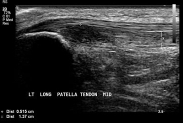
Ultrasound of the left patellar tendon demonstrates and area of decreased echotexture (dark area bordered by calipers) consistent with a large tear of the deep surface of the patellar tendon, commonly known as “Jumper’s knee”. (image 1)
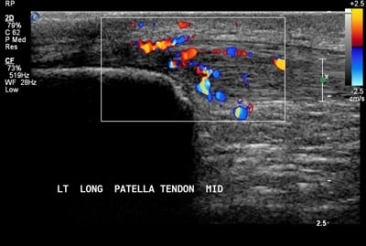
Colour flow demonstrates increased blood flow to the damaged tendon. (image 2)
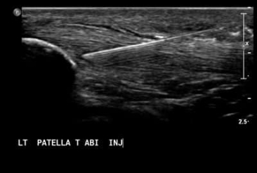
A needle is guided under ultrasound guidance directly to the area of maximal abnormality safely, ensuring accurate delivery of the patient’s blood. (image 3)
References:
- Creaney L, Hamilton B. Growth factor delivery methods in the management of sports injuries: the state of play. Br J Sports Med 42(5):314-20, 2008
- Edwards SG, Calandruccio JH. Autologous blood injections for refractory lateral epicondylitis. J Hand Surg [Am] 28(2):272-8, 2003
Further Information.
Referring doctors are welcome to discuss with our radiologists the imaging and interventional radiology needs of their patients and whether autologous blood injection is suitable for their patient’s medical condition.
