Ultrasound.
Fact Sheets | Diagnostic Imaging
Ultrasound.
Introduction
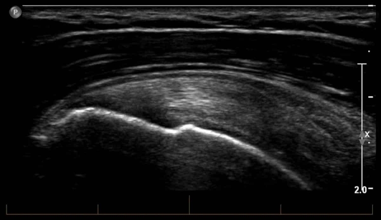
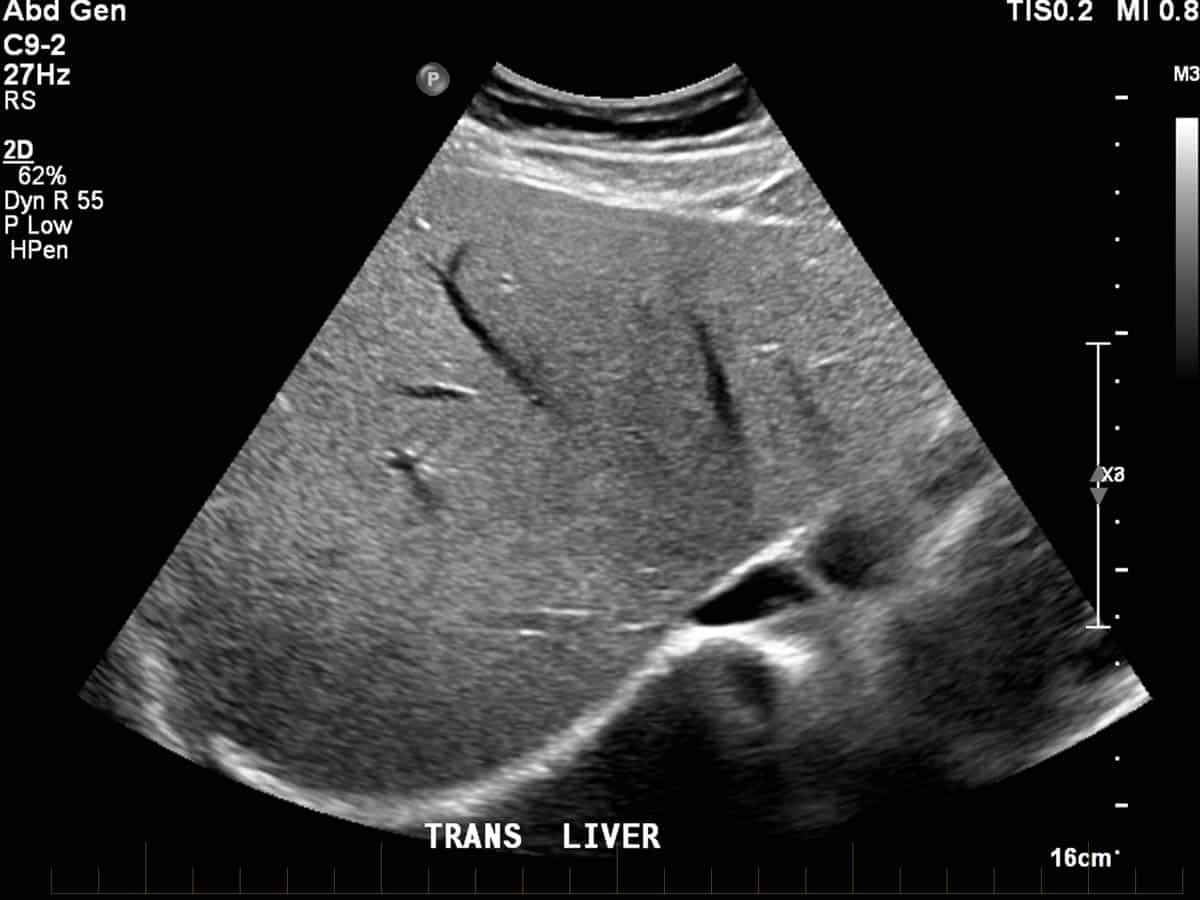
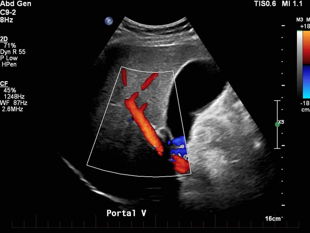
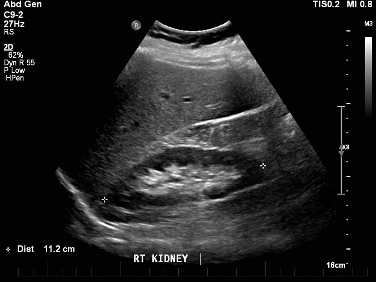
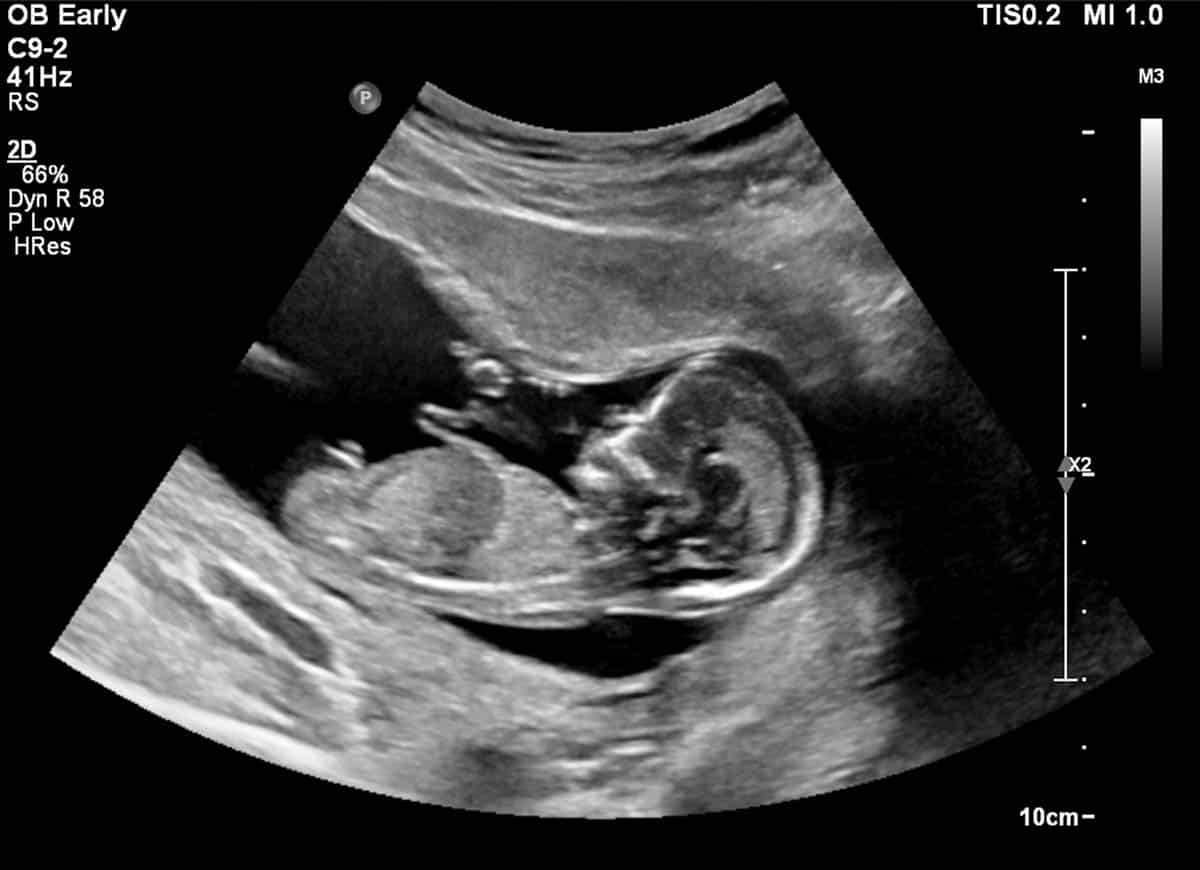
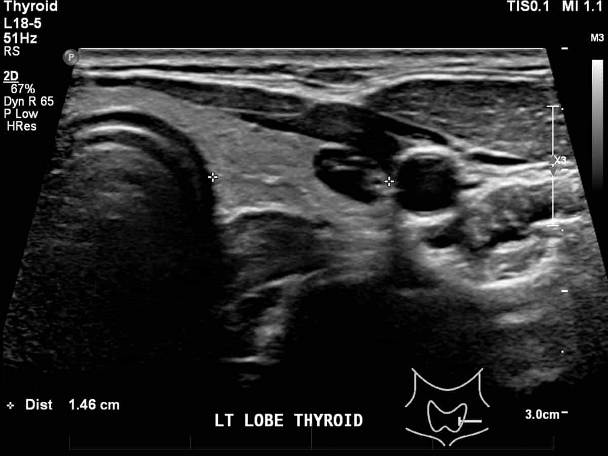
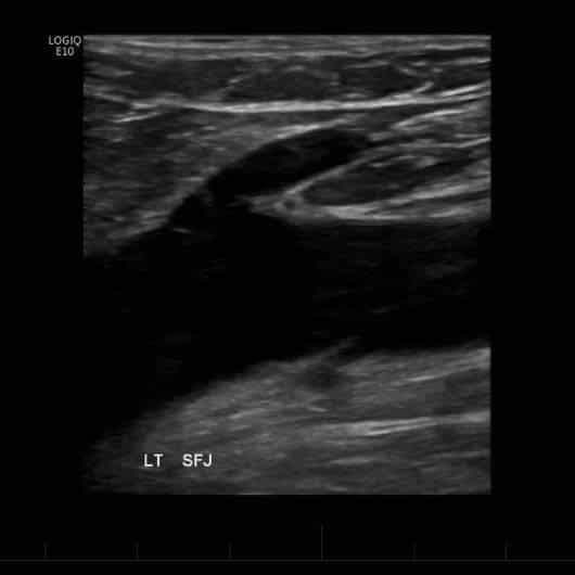
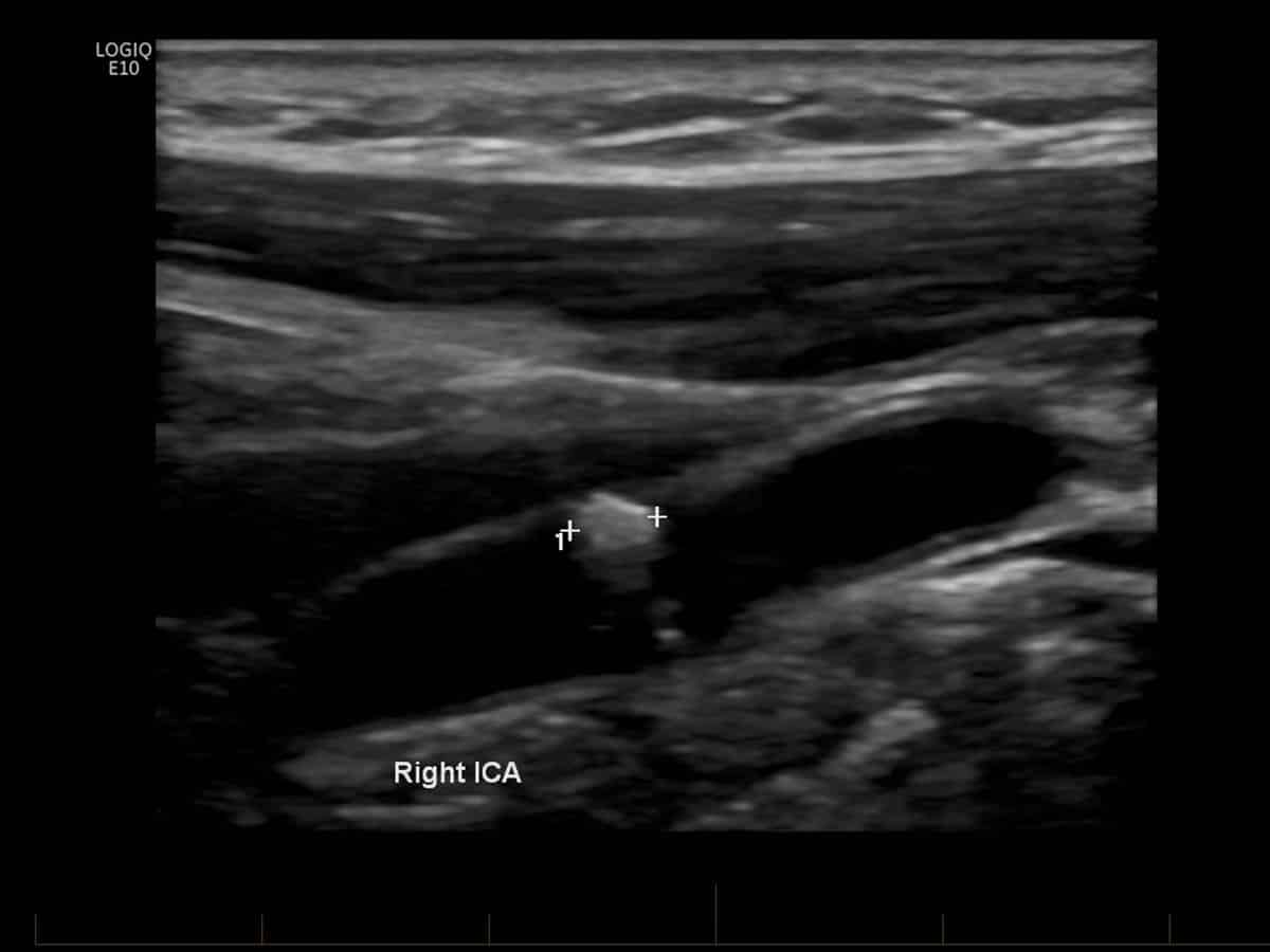
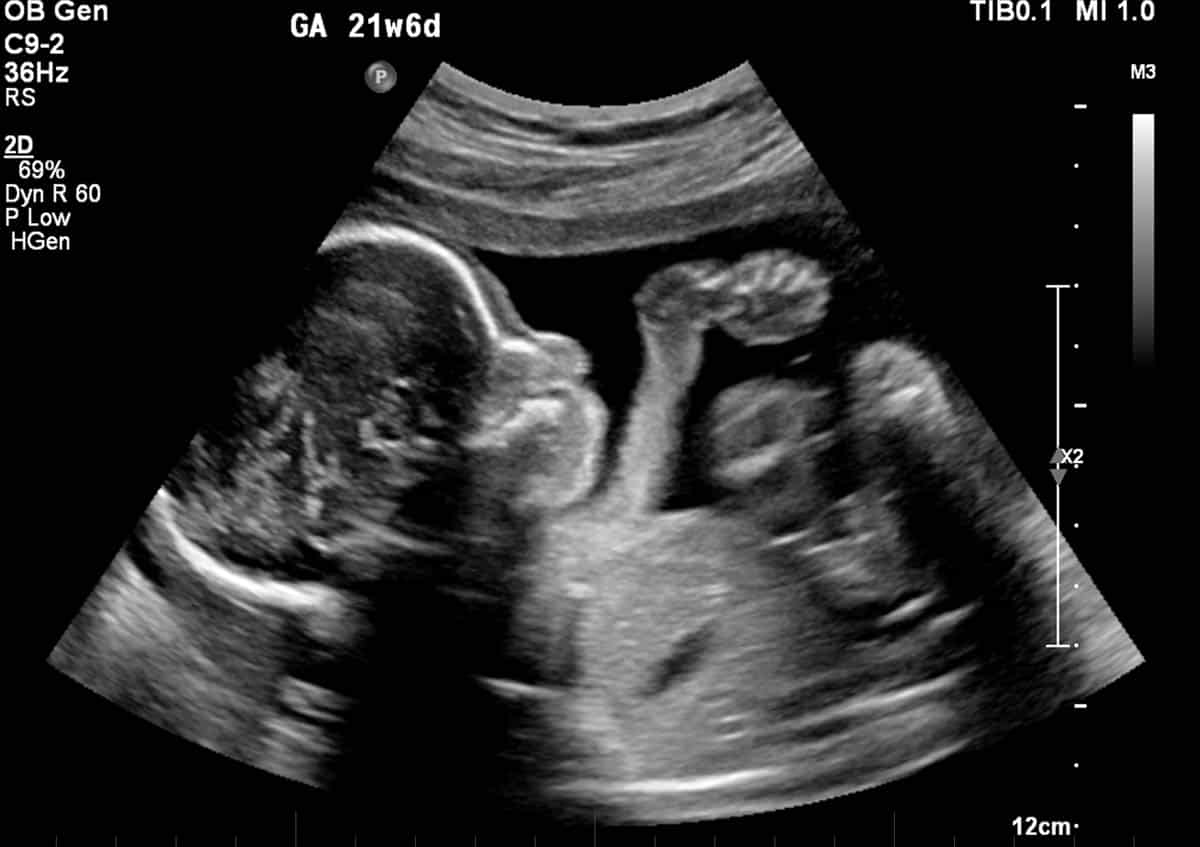
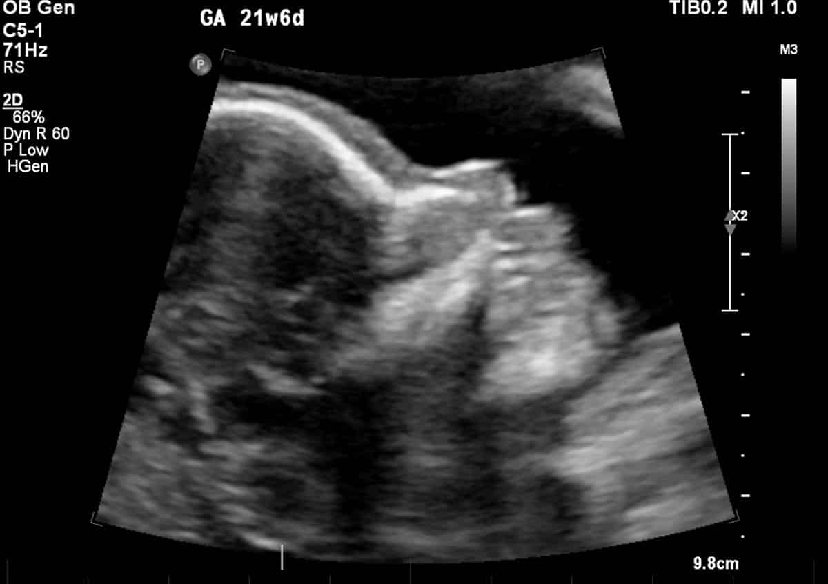
Ultrasound is a type of scan that uses inaudible high frequency sound waves to create detailed pictures of the body. It is a safe, painless and radiation free examination.
Ultrasound images can be taken during motion, so both still images and video images may be produced. The type of image provided to your doctor will depend upon what is requested, your individual circumstances and the preference of our doctor.
Preparation
Ultrasound examinations of the different body parts may require special preparation, such as fasting or drinking water.
Please take the time to read the summary of instructions below for the type of ultrasound examination you are to have. Our receptionist will provide you with the necessary instructions when you book your appointment and also answer any queries that you may have.
and Preparation Required
Fast for 6 hours prior (usually withhold breakfast).
Only have small sips of water for any medication that you may be taking
75 minutes prior to the appointment empty your bladder and then drink one litre of water (not coffee or tea), finishing the water at least one hour before the examination.
Do NOT empty your bladder until after the examination
Doppler veins - No preparation
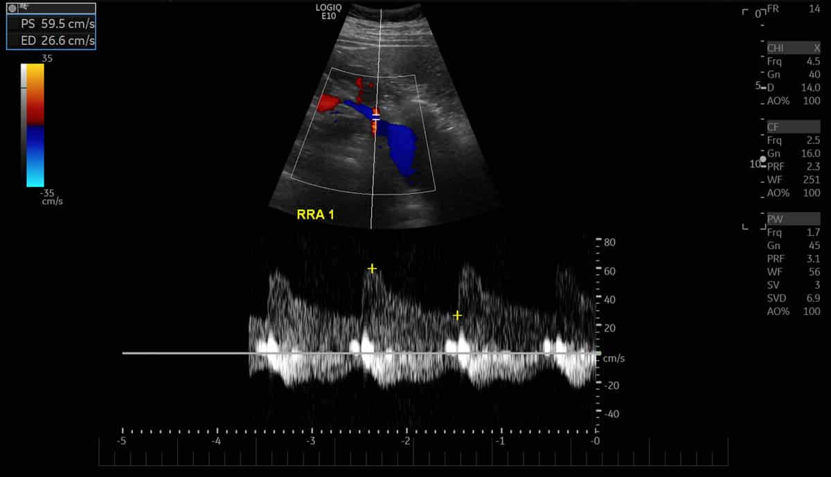
Doppler Carotid, Leg & other arteries - No preparation
Doppler Aorta & Renal - Fast 8 hours
No preparation
Procedure
Upon arrival at Melbourne Radiology Clinic, you will be taken to a change room. You will be requested to remove relevant clothing and jewellery and to wear the provided examination gown. The area to be examined will need to be exposed but the rest of you will be covered.
Procedures can take up to 1 hour to conduct with the average time being 20-30 minutes and will be discussed with you at the time of booking.
You will be asked to lie on a couch or sit on a chair (depending on the examination) next to the ultrasound equipment. A university-trained sonographer who is accredited with the Australian Sonographer Accreditation Registry (ASAR). captures and prints the requested images using a probe (also known as a transducer) which emits the ultrasound waves.
A water-based gel will be spread on the skin of the area being scanned which assists in the transmission of sound waves between the body and probe. The soundwaves are then reflected by the tissues in your body back to the probe. This information is then used to form an image that is used to obtain a diagnosis. You may be required to hold your breath or move into different positions so that the best images can be obtained.
Ultrasound waves cannot penetrate gas and bone. As such, ultrasound cannot be used to look beyond the soft tissue surrounding joints, into the lungs or gas containing bowel loops, however can obtain adequate images of solid organs such as the liver and uterus, as well as fluid filled organs such as the gall bladder.
Female Pelvic Ultrasound
The preparation requirements for female pelvic ultrasounds vary according to age, stage of pregnancy or type of condition requiring investigation.
- Women who are not pregnant and attending for a pelvic gynaecological scan need to have a full bladder, as do pregnant women in the first trimester (first three months).
- If you know that you are pregnant with twins, please let us know so that additional time may be allocated.
In addition, an internal or transvaginal examination may be requested by the referring doctor or required at the time of the examination for a closer view of pelvic organs, in particular the ovaries and endometrium (lining of the uterus). Even though an internal scan may be recommended, you naturally have a choice to refuse.
Results &
Follow-Up
One of Melbourne Radiology Clinic’s specialist radiologists, a medical doctor specialising in the interpretation of medical images for the purposes of providing a diagnosis, will then review the images and provide a formal written report. If medically urgent, or you have an appointment immediately after the scan to be seen by your doctor or health care provider, Melbourne Radiology Clinic will have your results ready without delay. Otherwise, the report will be received by your doctor or health care provider within the next 24 hours.
Please ensure that you make a follow up appointment with your referring doctor or health care provider to discuss your results.
Your referring doctor or health care provider is the most appropriate person to explain to you the results of the scans and for this reason, we do not release the results directly to you.
Reminders
 Digital X-Ray – Patient Fact Sheet
 Ultra-Low Dose CT Scan – Patient Fact Sheet
 Ultrasound – Patient Fact Sheet
 CT Intravenous Contrast & Consent – Patient Fact Sheet
 Magnetic Resonance Imaging (MRI) – Adult – Patient Guide
 Magnetic Resonance Imaging (MRI) – Children – Patient Guide
 What To Expect When Having A MRI – Video
 Radiation Safety – Patient Fact Sheet
